

Volume 36, Issue 257 (5-2026)
J Mazandaran Univ Med Sci 2026, 36(257): 3-15 |
Back to browse issues page



Download citation:
BibTeX | RIS | EndNote | Medlars | ProCite | Reference Manager | RefWorks
Send citation to:


BibTeX | RIS | EndNote | Medlars | ProCite | Reference Manager | RefWorks
Send citation to:
Shahi Y, Esmaeili-Bandboni A, Ghorbani-Anarkooli M, Mobayen M, Zaminy A, Asgari M. Protective Effects of Hemin on Adipose-Derived Mesenchymal Stem Cell Survival and Apoptosis under In Vitro Oxidative Stress. J Mazandaran Univ Med Sci 2026; 36 (257) :3-15
URL: http://jmums.mazums.ac.ir/article-1-22667-en.html
URL: http://jmums.mazums.ac.ir/article-1-22667-en.html
Yeganeh Shahi 
 , Aghil Esmaeili-Bandboni , Marjan Ghorbani-Anarkooli , Mohammadreza Mobayen , Arash Zaminy , Mehrdad Asgari *
, Aghil Esmaeili-Bandboni , Marjan Ghorbani-Anarkooli , Mohammadreza Mobayen , Arash Zaminy , Mehrdad Asgari *
Abstract: (257 Views)
Background and purpose: Oxidative stress is a major limiting factor affecting the survival of mesenchymal stem cells following transplantation. Activation of the heme oxygenase-1 (HMOX1) pathway is recognized as a key cytoprotective mechanism against oxidative damage. This study aimed to investigate the protective and anti-apoptotic effects of Hemin, a potent inducer of HMOX1, on human adipose-derived mesenchymal stem cells (hADSCs) exposed to hydrogen peroxide (H₂O₂)-induced oxidative stress.
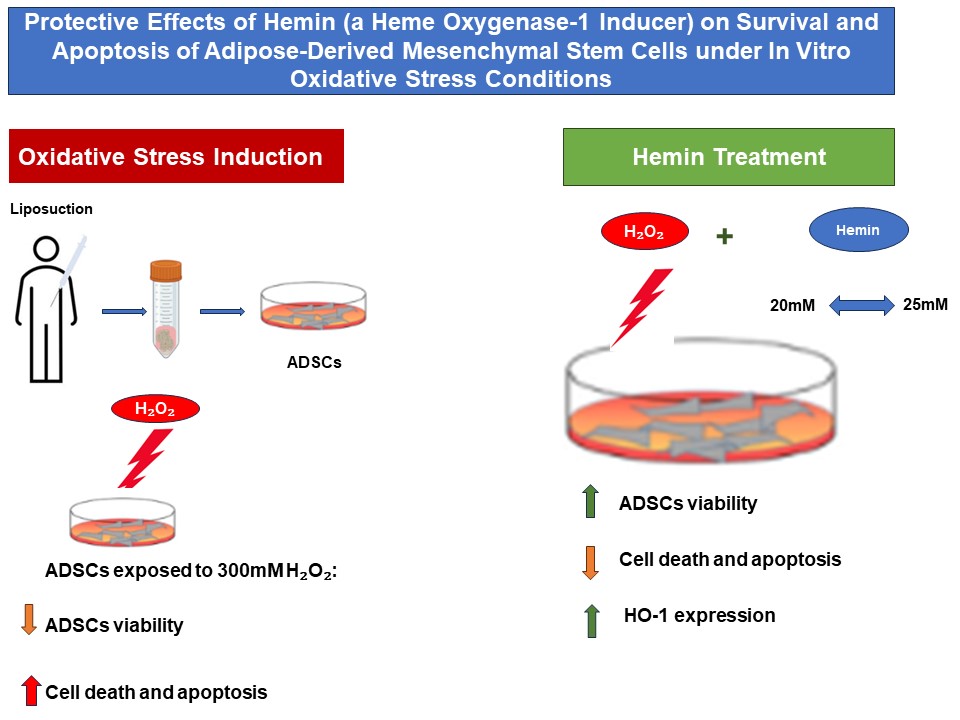
Materials and methods: hADSCs were isolated from human adipose tissue and characterized according to established mesenchymal stem cell markers. Cells were pretreated with non-cytotoxic concentrations of Hemin (10, 20, and 25 μM) and subsequently exposed to 300 μM H₂O₂, corresponding to the determined IC₅₀ value, for 24, 48, and 72 hours. Cell viability was assessed using the MTT assay, while apoptotic and morphological changes were evaluated using acridine orange/propidium iodide (AO/PI) and DAPI staining. The expression level of the HMOX1 gene was quantified using real-time RT-PCR.
Results: The MTT assay revealed that the IC₅₀ of H₂O₂ in hADSCs was 300 μM, whereas Hemin concentrations above 25 μM exhibited cytotoxic effects. Pretreatment with Hemin at 10, 20, and 25 μM significantly enhanced cell survival and attenuated H₂O₂-induced cytotoxicity at all evaluated time points. DAPI staining demonstrated a marked reduction in apoptotic cells, and AO/PI staining indicated improved cellular morphology and a decreased proportion of dead cells, particularly in the groups treated with 20 and 25 μM Hemin. The most pronounced protective effects were observed at the concentration of 25 μM after 48 and 72 hours. In addition, HMOX1 gene expression was significantly upregulated in Hemin-treated groups compared with the group exposed to oxidative stress alone.
Conclusion: Hemin enhances hADSC survival and reduces oxidative stress-induced apoptosis through the upregulation of HMOX1. These findings suggest that Hemin may serve as a promising cytoprotective agent for enhancing the efficacy of stem cell-based therapies.
Materials and methods: hADSCs were isolated from human adipose tissue and characterized according to established mesenchymal stem cell markers. Cells were pretreated with non-cytotoxic concentrations of Hemin (10, 20, and 25 μM) and subsequently exposed to 300 μM H₂O₂, corresponding to the determined IC₅₀ value, for 24, 48, and 72 hours. Cell viability was assessed using the MTT assay, while apoptotic and morphological changes were evaluated using acridine orange/propidium iodide (AO/PI) and DAPI staining. The expression level of the HMOX1 gene was quantified using real-time RT-PCR.
Results: The MTT assay revealed that the IC₅₀ of H₂O₂ in hADSCs was 300 μM, whereas Hemin concentrations above 25 μM exhibited cytotoxic effects. Pretreatment with Hemin at 10, 20, and 25 μM significantly enhanced cell survival and attenuated H₂O₂-induced cytotoxicity at all evaluated time points. DAPI staining demonstrated a marked reduction in apoptotic cells, and AO/PI staining indicated improved cellular morphology and a decreased proportion of dead cells, particularly in the groups treated with 20 and 25 μM Hemin. The most pronounced protective effects were observed at the concentration of 25 μM after 48 and 72 hours. In addition, HMOX1 gene expression was significantly upregulated in Hemin-treated groups compared with the group exposed to oxidative stress alone.
Conclusion: Hemin enhances hADSC survival and reduces oxidative stress-induced apoptosis through the upregulation of HMOX1. These findings suggest that Hemin may serve as a promising cytoprotective agent for enhancing the efficacy of stem cell-based therapies.
Keywords: Heme Oxygenase-1 (HMOX1), Human Adipose-Derived Mesenchymal Stem Cells (hADSCs), Oxidative Stress, Cytoprotection, Apoptosis.
Type of Study: Research(Original) |
Subject:
Biology
Send email to the article author
| Rights and permissions | |
 |
This work is licensed under a Creative Commons Attribution-NonCommercial 4.0 International License. |

Articles Copyright © The Author(s). Owned by Mazandaran University of Medical Sciences.
Contact Us
Address: Journal Office, Building No. 2, Mazandaran University of Medical Sciences, Moallem Square, Sari.Tel: +98 (011)34484874 Postal code: 48175-866 E-Mail: jmums@mazums.ac.ir Telefax: +98 (011)33262679
