

Volume 35, Issue 253 (1-2026)
J Mazandaran Univ Med Sci 2026, 35(253): 53-64 |
Back to browse issues page

![]()
![]()
![]()
Download citation:
BibTeX | RIS | EndNote | Medlars | ProCite | Reference Manager | RefWorks
Send citation to:


BibTeX | RIS | EndNote | Medlars | ProCite | Reference Manager | RefWorks
Send citation to:
Abouie Mehrizi O, Bodaghi Hosseinabadi R, Talebi A S. Dose Verification in Lung Radiotherapy Using PET Imaging of Nanoparticle-Induced Positrons: A Simulation Study with Bismuth and Hafnium Nanoparticles. J Mazandaran Univ Med Sci 2026; 35 (253) :53-64
URL: http://jmums.mazums.ac.ir/article-1-22555-en.html
URL: http://jmums.mazums.ac.ir/article-1-22555-en.html


Abstract: (715 Views)
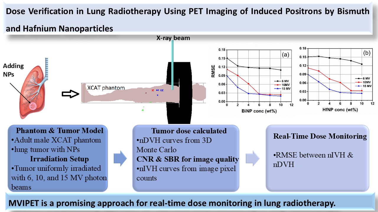
Background and purpose: Accurate verification of radiation dose delivery remains a major challenge in radiotherapy. Positron emission tomography imaging of megavoltage-induced positrons (MVIPET) has recently emerged as a potential in vivo dosimetry technique. This study investigated the feasibility of enhancing MVIPET signal intensity through the use of high-Z nanoparticles (NPs), specifically bismuth (Bi) and hafnium (Hf), to enable real-time dose monitoring during lung radiotherapy.
Materials and Methods: Positron emission tomography (PET) images resulting from positron emission induced by bismuth and silver nanoparticles within a lung tumour were generated using the GATE Monte Carlo simulation framework during irradiation with 6, 10, and 15 MV photon beams. The resulting images were assessed in terms of image quality and dose verification accuracy.
Results: Positron production, absorbed dose, and PET signal intensity increased with both photon beam energy and nanoparticle (NP) concentration, with bismuth nanoparticles (BiNPs) producing significantly greater enhancement than hafnium nanoparticles (HfNPs). High-quality MVIPET images with acceptable signal-to-background ratio (SBR) and contrast-to-noise ratio (CNR), as well as low root mean square error (RMSE), were achieved for BiNP concentrations ≥4 wt% at 10 MV and ≥2 wt% at 15 MV, with the minimum RMSE observed at 4 wt%. In contrast, HfNPs required higher concentrations to achieve reliable dose monitoring at both 10 and 15 MV photon energies. At 6 MV, image quality and dose–image correlation were insufficient to support clinical feasibility.
Conclusion: These findings indicate that MVIPET, when combined with bismuth and hafnium nanoparticles and higher photon beam energies (15 MV), represents a promising approach for real-time, non-invasive radiation dose verification in lung radiotherapy.
Type of Study: Research(Original) |
Subject:
Radiology
Send email to the article author
| Rights and permissions | |
 |
This work is licensed under a Creative Commons Attribution-NonCommercial 4.0 International License. |

Articles Copyright © The Author(s). Owned by Mazandaran University of Medical Sciences.
Contact Us
Address: Journal Office, Building No. 2, Mazandaran University of Medical Sciences, Moallem Square, Sari.Tel: +98 (011)34484874 Postal code: 48175-866 E-Mail: jmums@mazums.ac.ir Telefax: +98 (011)33262679
