

Volume 34, Issue 242 (2-2025)
J Mazandaran Univ Med Sci 2025, 34(242): 59-67 |
Back to browse issues page

![]()
![]()
![]()
Download citation:
BibTeX | RIS | EndNote | Medlars | ProCite | Reference Manager | RefWorks
Send citation to:


BibTeX | RIS | EndNote | Medlars | ProCite | Reference Manager | RefWorks
Send citation to:
Jafari N, Mahmoudi S M, Taherneghad F. Mast Cell Frequency in Oral Squamous Cell Carcinoma. J Mazandaran Univ Med Sci 2025; 34 (242) :59-67
URL: http://jmums.mazums.ac.ir/article-1-21433-en.html
URL: http://jmums.mazums.ac.ir/article-1-21433-en.html


Abstract: (1354 Views)
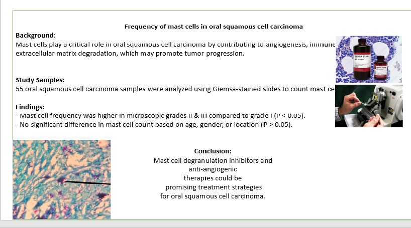
Background and purpose: Oral squamous cell carcinoma is the most common malignancy of the oral cavity. In solid tumors, cancer cells and stromal cells (fibroblasts, inflammatory cells, endothelial cells) play a role in tumor progression, angiogenesis, local invasion, and metastasis. Mast cells contribute to carcinogenesis by releasing chemical mediators in their granules through various pathways, including suppressing the immune system, enhancing angiogenesis, destroying the extracellular matrix, and increasing the mitosis of tumoral cells. Several studies have investigated the role of mast cells in oral squamous cell carcinoma. While some suggest that mast cells play an angiogenic and tumorigenic role, others do not confirm this, highlighting the contradictory findings in this area. In this study, we discuss the role of mast cells in oral squamous cell carcinoma.
Materials and methods: The mean number of mast cells was significantly lower in higher microscopic grades compared to poor grades (P<0.05). However, there was no significant difference in the average number of mast cells based on age, gender, or location of occurrence (P>0.05).
Results: the mean number of mast cells was significantly lower in microscopic grade and higher than poor (P-value˂0/05). But there was no significant difference in the average number of mast cells based on age, gender and place of occurrence (P˃0/05).
Conclusion: The significant increase in the average number of mast cells in microscopic grades II and III compared to grade I indicates the role of mast cells as an indicator of disease progression. This finding may support the use of novel treatment approaches, such as mast cell degranulation inhibitors and anti-angiogenic therapies.
Type of Study: Research(Original) |
Subject:
Pathology
Send email to the article author
| Rights and permissions | |
 |
This work is licensed under a Creative Commons Attribution-NonCommercial 4.0 International License. |

Articles Copyright © The Author(s). Owned by Mazandaran University of Medical Sciences.
Contact Us
Address: Journal Office, Building No. 2, Mazandaran University of Medical Sciences, Moallem Square, Sari.Tel: +98 (011)34484874 Postal code: 48175-866 E-Mail: jmums@mazums.ac.ir Telefax: +98 (011)33262679
