

Volume 36, Issue 263 (11-2026)
J Mazandaran Univ Med Sci 2026, 36(263): 135-145 |
Back to browse issues page



Download citation:
BibTeX | RIS | EndNote | Medlars | ProCite | Reference Manager | RefWorks
Send citation to:


BibTeX | RIS | EndNote | Medlars | ProCite | Reference Manager | RefWorks
Send citation to:
Zamanzadeh M, Shiva A, Hamidzadeh F, Hosseinnataj A, Koochak Dezfouli M. Inflammation Severity and Histopathological Features in Radicular Cysts: Frequency and Associations with Age and Sex. , J Mazandaran Univ Med Sci. 2026; 36 (263) :135-145
URL: http://jmums.mazums.ac.ir/article-1-23097-en.html
URL: http://jmums.mazums.ac.ir/article-1-23097-en.html
Maryam Zamanzadeh 
 , Atena Shiva , Faezeh Hamidzadeh , Abolfazl Hosseinnataj , Mohammad Koochak Dezfouli *
Abstract: (15 Views)
Background and purpose: Radicular cysts are the most common inflammatory odontogenic cysts of the jaws and develop following pulpal inflammation and necrosis, which stimulate the proliferation of epithelial remnants. These lesions exhibit a range of inflammatory and histopathological changes. Assessing inflammatory severity alongside histopathological features may provide further insight into the tissue changes associated with these lesions. This study aimed to determine the frequency of histopathological features in radicular cysts and examine their associations with inflammation severity, age, and sex.
Materials and methods: This retrospective cross-sectional study included 126 paraffin-embedded tissue blocks with histopathologically confirmed diagnoses of radicular cysts from a pathology laboratory in Sari, Iran. Data on age, sex, and lesion location were obtained from patient records. Haematoxylin and eosin (H&E)-stained sections were examined by two oral and maxillofacial pathologists. Epithelial thickness, the severity and type of inflammation, and the presence of histopathological features were recorded. Associations were analysed using the chi-square test or Fisher’s exact test, as appropriate. Statistical significance was set at P < 0.05.
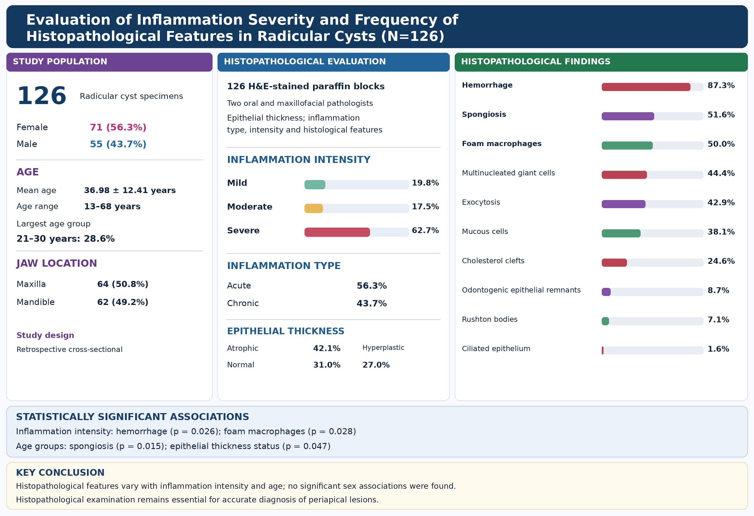
Results: Of the 126 specimens, 71 (56.3%) were from female patients and 55 (43.7%) from male patients. The mean age was 36.98 ± 12.41 years. Severe inflammation was observed in 62.7% of specimens. The most frequent histopathological findings were haemorrhage (87.3%), spongiosis (51.6%), foamy macrophages (50.0%), and multinucleated giant cells (44.4%). Haemorrhage (P = 0.026) and foamy macrophages (P = 0.028) were significantly associated with inflammation severity. Spongiosis (P = 0.015) and epithelial thickness category (P = 0.047) were significantly associated with age group. No significant associations were found between the assessed histopathological features and sex.
Conclusion: Radicular cysts exhibited a diverse range of histopathological features. Haemorrhage and foamy macrophages were associated with inflammation severity, whereas spongiosis and epithelial thickness category were associated with age group. These findings highlight the histopathological variability of radicular cysts and support the value of histopathological examination in their assessment and differentiation from other periapical lesions.
Materials and methods: This retrospective cross-sectional study included 126 paraffin-embedded tissue blocks with histopathologically confirmed diagnoses of radicular cysts from a pathology laboratory in Sari, Iran. Data on age, sex, and lesion location were obtained from patient records. Haematoxylin and eosin (H&E)-stained sections were examined by two oral and maxillofacial pathologists. Epithelial thickness, the severity and type of inflammation, and the presence of histopathological features were recorded. Associations were analysed using the chi-square test or Fisher’s exact test, as appropriate. Statistical significance was set at P < 0.05.
Results: Of the 126 specimens, 71 (56.3%) were from female patients and 55 (43.7%) from male patients. The mean age was 36.98 ± 12.41 years. Severe inflammation was observed in 62.7% of specimens. The most frequent histopathological findings were haemorrhage (87.3%), spongiosis (51.6%), foamy macrophages (50.0%), and multinucleated giant cells (44.4%). Haemorrhage (P = 0.026) and foamy macrophages (P = 0.028) were significantly associated with inflammation severity. Spongiosis (P = 0.015) and epithelial thickness category (P = 0.047) were significantly associated with age group. No significant associations were found between the assessed histopathological features and sex.
Conclusion: Radicular cysts exhibited a diverse range of histopathological features. Haemorrhage and foamy macrophages were associated with inflammation severity, whereas spongiosis and epithelial thickness category were associated with age group. These findings highlight the histopathological variability of radicular cysts and support the value of histopathological examination in their assessment and differentiation from other periapical lesions.
Type of Study: Research(Original) |
Subject:
Dentistry
Send email to the article author
| Rights and permissions | |
 |
This work is licensed under a Creative Commons Attribution-NonCommercial 4.0 International License. |

Articles Copyright © The Author(s). Owned by Mazandaran University of Medical Sciences.
Contact Us
Address: Journal Office, Building No. 2, Mazandaran University of Medical Sciences, Moallem Square, Sari.Tel: +98 (011)34484874 Postal code: 48175-866 E-Mail: jmums@mazums.ac.ir Telefax: +98 (011)33262679
