

Volume 36, Issue 259 (8-2026)
J Mazandaran Univ Med Sci 2026, 36(259): 179-183 |
Back to browse issues page



Download citation:
BibTeX | RIS | EndNote | Medlars | ProCite | Reference Manager | RefWorks
Send citation to:


BibTeX | RIS | EndNote | Medlars | ProCite | Reference Manager | RefWorks
Send citation to:
Mohammadnejad F, Golchinmehr S. A Rare Diagnosis: A Case of Mounier-Kuhn Syndrome. J Mazandaran Univ Med Sci 2026; 36 (259) :179-183
URL: http://jmums.mazums.ac.ir/article-1-22616-en.html
URL: http://jmums.mazums.ac.ir/article-1-22616-en.html


Abstract: (543 Views)
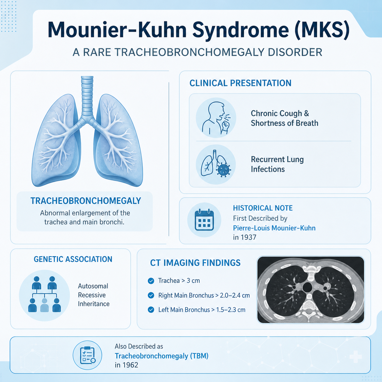
Mounier-Kuhn syndrome (MKS) is a rare congenital disorder characterized by dilatation of the trachea and main bronchi. It was first described by Pierre-Louis Mounier-Kuhn in 1937 in association with recurrent respiratory infections and was later anatomically described as tracheobronchomegaly (TBM) in 1962. The aetiology of Mounier-Kuhn syndrome is unknown, but familial predisposition and autosomal recessive inheritance are thought to play a role in its development. Patients usually present with chronic cough and shortness of breath, which may resemble the symptoms of chronic obstructive pulmonary disease. Radiographic evidence of abnormal dilatation of the trachea and main bronchi may be seen. On CT imaging, Mounier-Kuhn syndrome should be suspected when the diameters of the trachea, right main bronchus, and left main bronchus exceed 3.0 cm, 2.4 cm, and 2.3 cm, respectively. Complications associated with MKS include bronchiectasis and emphysema, among others. Given its rarity, there is no standard protocol for the management of this syndrome. Our patient, a 46-year-old non-smoking man with no previous medical history, was admitted to the hospital following cardiorespiratory arrest. During the treatment process, the patient developed biliary reflux, and a bag connected to a gastric tube was inserted due to a suspected tracheoesophageal fistula. CT revealed a main bronchial diameter of more than 32 mm. Finally, during investigations conducted in the intensive care unit, findings consistent with Mounier-Kuhn syndrome were identified on examination and CT imaging. The findings are discussed in this article.
Type of Study: Case Report |
Subject:
genetic
Send email to the article author
| Rights and permissions | |
 |
This work is licensed under a Creative Commons Attribution-NonCommercial 4.0 International License. |

Articles Copyright © The Author(s). Owned by Mazandaran University of Medical Sciences.
Contact Us
Address: Journal Office, Building No. 2, Mazandaran University of Medical Sciences, Moallem Square, Sari.Tel: +98 (011)34484874 Postal code: 48175-866 E-Mail: jmums@mazums.ac.ir Telefax: +98 (011)33262679
