

Volume 36, Issue 259 (8-2026)
J Mazandaran Univ Med Sci 2026, 36(259): 184-190 |
Back to browse issues page



Download citation:
BibTeX | RIS | EndNote | Medlars | ProCite | Reference Manager | RefWorks
Send citation to:


BibTeX | RIS | EndNote | Medlars | ProCite | Reference Manager | RefWorks
Send citation to:
Aliabadi T, Eghtesad M A, Khatibi S F. Surgical Extrusion as an Alternative Treatment Approach for the Restoration of Teeth with Subgingival Structural Defects: A Case Series. J Mazandaran Univ Med Sci 2026; 36 (259) :184-190
URL: http://jmums.mazums.ac.ir/article-1-22774-en.html
URL: http://jmums.mazums.ac.ir/article-1-22774-en.html


Abstract: (558 Views)
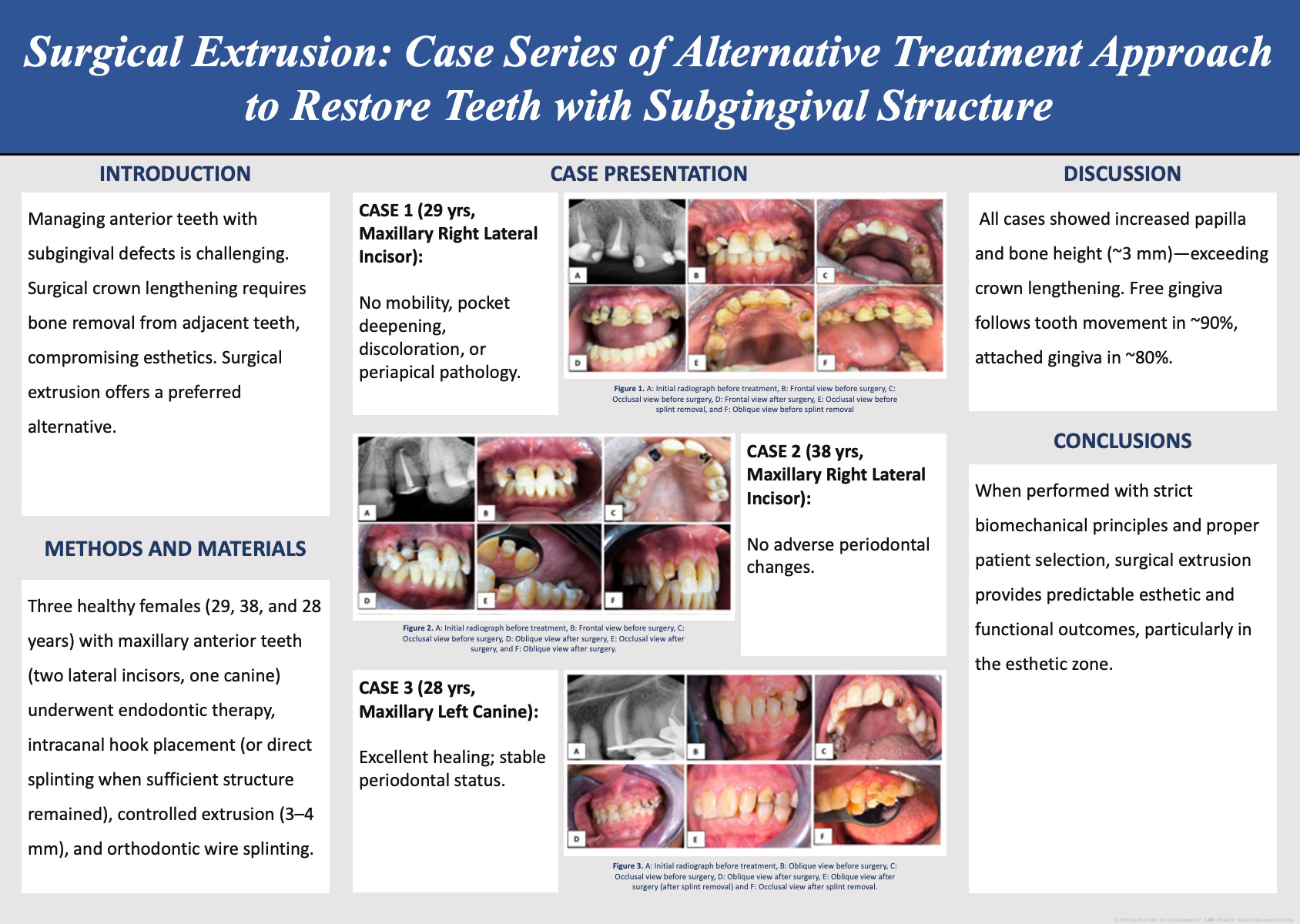
Surgical extrusion represents an effective therapeutic approach for the rehabilitation of teeth with subgingival structural defects while preserving root structure in the esthetic zone. Managing and restoring single teeth positioned apically to the alveolar bone crest presents significant clinical challenges, particularly when associated with caries, crown fractures, or external root resorption. Surgical crown lengthening, particularly for maxillary anterior teeth, may result in esthetic complications because establishing adequate biological width often requires osseous removal involving adjacent teeth. Therefore, surgical extrusion has emerged as an alternative tooth-preservation strategy in the esthetic zone. This case series describes three systemically healthy females (aged 28, 29, and 38 years) with maxillary anterior teeth (two lateral incisors and one canine) exhibiting subgingival structural defects. The patients underwent endodontic therapy, intracanal hook placement using resin-modified glass ionomer (RMGI) cement, controlled surgical extrusion of 3-4 mm using elevators and forceps, and immediate stabilization with a 0.5-mm orthodontic wire and flowable composite resin. In cases with sufficient supragingival tooth structure, direct composite splinting was performed. Clinical and radiographic assessments were conducted at 48 hours, 2 weeks (splint removal), and 6 weeks (prosthetic restoration). Case 1 (29-year-old patient with a maxillary right lateral incisor) showed no adverse periodontal findings, including mobility, increased probing depth, gingival discoloration, or periapical pathology. Case 2 (38-year-old patient with a maxillary right lateral incisor) demonstrated stable periodontal conditions without adverse soft tissue changes. Case 3 (28-year-old patient with a maxillary left canine) demonstrated successful extrusion with favourable healing and periodontal stability. All cases showed approximately 3-mm increases in interdental papilla height and alveolar bone crest height. Within the limitations of this case series, surgical extrusion performed according to appropriate biomechanical principles and patient selection criteria appears to be a predictable approach for preserving structurally compromised teeth and achieving favourable esthetic and functional outcomes.
Type of Study: Case Report |
Subject:
Dentistry
Send email to the article author
| Rights and permissions | |
 |
This work is licensed under a Creative Commons Attribution-NonCommercial 4.0 International License. |

Articles Copyright © The Author(s). Owned by Mazandaran University of Medical Sciences.
Contact Us
Address: Journal Office, Building No. 2, Mazandaran University of Medical Sciences, Moallem Square, Sari.Tel: +98 (011)34484874 Postal code: 48175-866 E-Mail: jmums@mazums.ac.ir Telefax: +98 (011)33262679
