

Volume 36, Issue 263 (11-2026)
J Mazandaran Univ Med Sci 2026, 36(263): 238-245 |
Back to browse issues page



Download citation:
BibTeX | RIS | EndNote | Medlars | ProCite | Reference Manager | RefWorks
Send citation to:


BibTeX | RIS | EndNote | Medlars | ProCite | Reference Manager | RefWorks
Send citation to:
Mobarakshahi A, Hamidian A H, Mehrabadifard F, Nasiri E. Pons Hepatis Forming a Tunnel for the Ligamentum Teres Hepatis: A Rare Anatomical Case Report. , J Mazandaran Univ Med Sci. 2026; 36 (263) :238-245
URL: http://jmums.mazums.ac.ir/article-1-23144-en.html
URL: http://jmums.mazums.ac.ir/article-1-23144-en.html


Abstract: (35 Views)
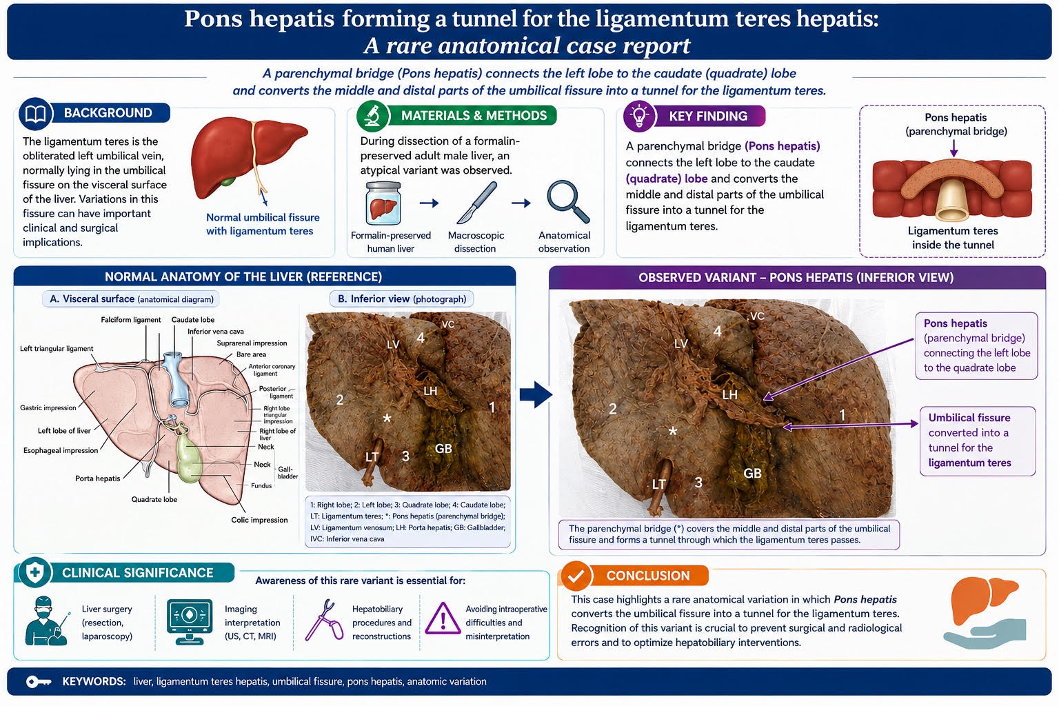
Background and purpose: The umbilical fissure (UF), which contains the ligamentum teres hepatis (LTH), is an important anatomical landmark of the liver. The LTH represents the embryonic remnant of the left umbilical vein (LUV). The UF is an important anatomical landmark for surgeons performing hepatic resections, radiologists interpreting liver imaging, and anatomists investigating hepatic morphology. Anatomical variations in this region, including the presence of a pons hepatis (PH) and morphological alterations of adjacent liver lobes, may have important clinical implications for the diagnosis and management of hepatic disorders.
Materials and methods: This case report was prepared following approval by the Ethics Committee of Guilan University of Medical Sciences, Rasht, Iran. The specimen was a formalin-fixed human liver obtained from an adult male cadaver of Iranian origin, whose age was unknown, and used for gross anatomy teaching at Guilan University of Medical Sciences. On gross examination, the liver appeared normal, with no visible pathological changes.
Results: In the examined specimen, the caudate lobe (CL) and quadrate lobe (QL) showed unusual morphological configurations compared with the typical anatomical arrangement. A PH of variable dimensions connected the QL to the left lobe (LL). This parenchymal bridge was located deep within the UF and covered its upper third. The presence of the PH resulted in a fusion between the LL and QL, converting the middle and distal portions of the UF into a tunnel through which the LTH passed.
Conclusion: The presence of a PH within the UF, together with associated morphological variations of the caudate and quadrate lobes, represents a rare anatomical variant. Awareness of this variation is important when interpreting liver imaging and planning abdominal surgical procedures, particularly hepatic, gallbladder, and biliary tract surgeries, to minimise the risk of inadvertent injury to adjacent anatomical structures.
Materials and methods: This case report was prepared following approval by the Ethics Committee of Guilan University of Medical Sciences, Rasht, Iran. The specimen was a formalin-fixed human liver obtained from an adult male cadaver of Iranian origin, whose age was unknown, and used for gross anatomy teaching at Guilan University of Medical Sciences. On gross examination, the liver appeared normal, with no visible pathological changes.
Results: In the examined specimen, the caudate lobe (CL) and quadrate lobe (QL) showed unusual morphological configurations compared with the typical anatomical arrangement. A PH of variable dimensions connected the QL to the left lobe (LL). This parenchymal bridge was located deep within the UF and covered its upper third. The presence of the PH resulted in a fusion between the LL and QL, converting the middle and distal portions of the UF into a tunnel through which the LTH passed.
Conclusion: The presence of a PH within the UF, together with associated morphological variations of the caudate and quadrate lobes, represents a rare anatomical variant. Awareness of this variation is important when interpreting liver imaging and planning abdominal surgical procedures, particularly hepatic, gallbladder, and biliary tract surgeries, to minimise the risk of inadvertent injury to adjacent anatomical structures.
Type of Study: Case Report |
Subject:
anatomy
| Rights and permissions | |
 |
This work is licensed under a Creative Commons Attribution-NonCommercial 4.0 International License. |

Articles Copyright © The Author(s). Owned by Mazandaran University of Medical Sciences.
Contact Us
Address: Journal Office, Building No. 2, Mazandaran University of Medical Sciences, Moallem Square, Sari.Tel: +98 (011)34484874 Postal code: 48175-866 E-Mail: jmums@mazums.ac.ir Telefax: +98 (011)33262679
